One of the biggest concerns for pet parents isn’t whether their veterinarian recommends an ultrasound—it’s understanding how much the procedure will actually cost. Unlike a routine consultation, ultrasound pricing can vary depending on the type of scan, the clinic’s facilities, whether a veterinary radiologist is involved, and whether additional diagnostics are required.
In Gurgaon, most veterinary clinics and multispecialty pet hospitals offer ultrasound as part of their diagnostic services. A basic abdominal ultrasound generally costs less than a specialised cardiac ultrasound or an emergency FAST scan performed during critical care. If sedation, blood tests, or ultrasound-guided procedures are recommended, the final bill may increase accordingly.
The good news is that an ultrasound remains one of the most cost-effective diagnostic tools available. It provides detailed images of internal organs without surgery or radiation, helping veterinarians diagnose problems accurately and begin treatment sooner.
Instead of focusing only on the lowest price, it’s important to understand what is included in the quoted fee. A clinic that charges slightly more may include specialist interpretation, digital reports, and advanced imaging equipment, ultimately providing greater value and a more accurate diagnosis.
Pet Ultrasound Cost in Gurgaon (2026)

Why Do Ultrasound Prices Vary So Much?
If you’ve contacted multiple veterinary clinics, you’ve probably noticed that the prices can differ significantly. This doesn’t necessarily mean one clinic is overcharging. In most cases, the difference reflects the level of expertise, technology, and services included in the examination.
For example, a neighbourhood veterinary clinic may perform a routine abdominal ultrasound at a lower cost, while a multispecialty pet hospital with advanced imaging equipment and a dedicated radiologist may charge more. Emergency scans performed late at night or on holidays also tend to cost more because they require immediate specialist attention.
The complexity of your pet’s condition also matters. A simple pregnancy confirmation usually takes less time than a detailed abdominal scan investigating liver disease, kidney problems, or a suspected tumour. Some pets remain calm throughout the procedure, while others may require mild sedation for accurate imaging, adding to the overall expense.

Factors That Influence Ultrasound Costs
| Factor | Impact on Cost |
|---|---|
| Type of Ultrasound | High |
| Veterinary Specialist | High |
| Emergency Appointment | High |
| Sedation (if required) | Moderate |
| Blood Tests Before Scan | Moderate |
| Ultrasound-Guided Procedures | High |
| Clinic Infrastructure | Moderate |
| Report by Radiologist | Moderate |
What Does a Pet Ultrasound Actually Show?
Many pet owners assume that an ultrasound is only used during pregnancy. In reality, it is one of the most versatile diagnostic tools available in veterinary medicine.
Unlike X-rays, which primarily show bones and dense structures, ultrasound allows veterinarians to examine soft tissues in real time. They can evaluate how organs move, measure blood flow, detect abnormal fluid accumulation, identify tumours, monitor pregnancies, and assess internal injuries without making a single incision.
This makes ultrasound invaluable for diagnosing conditions that would otherwise remain hidden during a routine physical examination.
For dogs and cats experiencing unexplained vomiting, persistent diarrhoea, abdominal swelling, urinary problems, or weight loss, an ultrasound often provides answers that blood tests alone cannot.
Common Conditions Diagnosed Using Ultrasound

When Does Your Dog or Cat Need an Ultrasound?
Not every illness requires advanced imaging, but there are situations where an ultrasound becomes one of the most valuable diagnostic tools available. Veterinarians typically recommend it when symptoms suggest that an internal organ may be affected or when routine blood tests and physical examinations do not provide a clear diagnosis.
A dog that has been vomiting for several days, a cat with recurring urinary issues, or a pregnant pet needing fetal monitoring are all common examples where an ultrasound can provide crucial information. It can also help evaluate liver enlargement, kidney disease, pancreatitis, abdominal masses, internal bleeding, reproductive disorders, and unexplained weight loss.
In emergency situations, veterinarians often perform a FAST (Focused Assessment with Sonography for Trauma) ultrasound to quickly check for internal bleeding or fluid accumulation after accidents or severe trauma.
Rather than replacing other diagnostic tests, ultrasound works alongside blood tests, X-rays, and physical examinations to give veterinarians a more complete understanding of your pet’s health.
Ultrasound vs X-ray: Which One Is Better?
One of the most common questions pet owners ask is whether an ultrasound is better than an X-ray. The truth is that neither test is universally better—they simply serve different purposes.
An X-ray excels at showing bones, fractures, chest conditions, swallowed metal objects, and certain lung diseases. Ultrasound, on the other hand, provides detailed images of soft tissues such as the liver, kidneys, bladder, intestines, pancreas, spleen, and reproductive organs.
Many veterinarians actually recommend both tests together because they complement each other. For example, a dog with persistent vomiting may first undergo an X-ray to check for obvious obstructions. If the cause remains unclear, an ultrasound can provide a much closer look at the stomach, intestines, and surrounding organs.
How to Prepare Your Dog or Cat Before an Ultrasound
If your veterinarian has scheduled an ultrasound, you might be wondering whether there’s anything you need to do before bringing your pet to the clinic. The answer is yes. A little preparation can make the examination faster, more comfortable, and more accurate.
In many cases, veterinarians recommend withholding food for 8 to 12 hours before an abdominal ultrasound. An empty stomach reduces the amount of gas in the digestive tract, allowing the ultrasound waves to produce clearer images of organs such as the liver, pancreas, kidneys, intestines, and bladder. Water is usually allowed unless your veterinarian advises otherwise.
Another important step is ensuring your pet has a moderately full bladder. For urinary tract examinations, a partially filled bladder provides much better visualization than an empty one. Your veterinarian may ask you to avoid allowing your dog or cat to urinate immediately before the appointment.
It’s also a good idea to carry previous blood test reports, X-rays, prescriptions, and medical records. These help the veterinarian compare findings and understand your pet’s medical history.
If your pet is particularly anxious or aggressive, inform the clinic in advance. While most dogs and cats tolerate ultrasound very well, some may require mild sedation to remain still during the examination.
Preparing Your Pet for an Ultrasound
| Preparation | Recommendation |
|---|---|
| Food | Fast for 8–12 hours (if advised) |
| Water | Usually allowed unless instructed otherwise |
| Bladder | Keep moderately full for urinary scans |
| Medical Records | Carry previous reports and prescriptions |
| Medications | Inform the veterinarian about ongoing medicines |
| Leash/Carrier | Essential for safe transportation |
| Nervous Pets | Discuss sedation options beforehand |

What Happens During a Pet Ultrasound?
One reason many pet owners feel anxious is simply because they don’t know what happens during the procedure. Fortunately, a veterinary ultrasound is completely non-invasive and is generally well tolerated by both dogs and cats.
Once your pet arrives at the clinic, the veterinarian performs a brief physical examination and reviews the medical history. If an abdominal ultrasound has been recommended, a small area of fur on the abdomen may be clipped. This allows the ultrasound probe to make direct contact with the skin and improves image quality.
A special ultrasound gel is then applied to the skin. This gel helps transmit sound waves efficiently and is completely safe for pets.
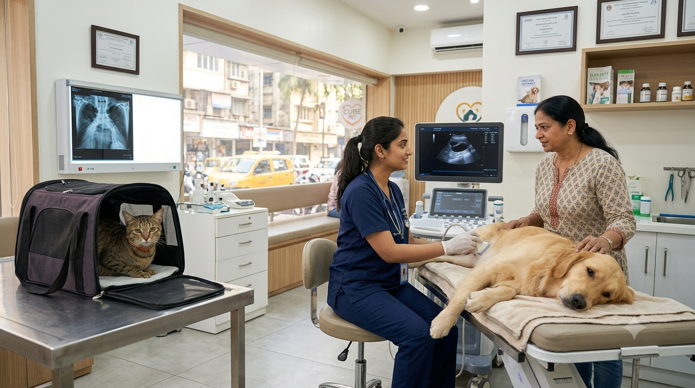
Your dog or cat is usually asked to lie comfortably on a padded examination table while the veterinarian gently moves the ultrasound probe over the area being examined. Throughout the procedure, images appear instantly on a monitor, allowing the veterinarian to evaluate internal organs in real time.
Unlike X-rays, ultrasound doesn’t just capture a single picture. It allows the veterinarian to observe organs as they function, monitor blood flow, identify abnormal fluid, measure masses, and assess the structure of tissues.
In most cases, the scan takes between 20 and 40 minutes, depending on the complexity of the examination.
Step-by-Step Ultrasound Procedure
| Step | What Happens |
|---|---|
| Appointment & Registration | Medical history and symptoms reviewed |
| Physical Examination | Initial assessment by veterinarian |
| Preparation | Hair clipping and ultrasound gel application |
| Ultrasound Scan | Real-time imaging of internal organs |
| Image Interpretation | Veterinarian evaluates findings |
| Report Discussion | Results explained to the pet owner |
| Treatment Plan | Further tests or treatment recommended if necessary |
Is an Ultrasound Painful for Dogs or Cats?
This is one of the most common questions veterinarians receive, and thankfully, the answer is reassuring.
An ultrasound is not painful. Unlike surgery or invasive diagnostic procedures, it does not involve needles, incisions, or radiation. The probe simply glides over your pet’s skin while emitting harmless sound waves that create images of internal organs.
Most pets remain calm throughout the examination, especially if they are accustomed to veterinary visits. Some may feel slightly uncomfortable if the veterinarian needs to apply gentle pressure over a painful area, but the procedure itself does not cause pain.
In fact, many dogs become so relaxed during the examination that they lie quietly for the entire scan.
Will My Pet Need Sedation?
The vast majority of ultrasound examinations are performed without sedation.
However, there are certain situations where mild sedation may be recommended. Pets that are extremely anxious, aggressive, or unable to remain still may require sedation to ensure accurate imaging. Likewise, animals experiencing severe pain may be more comfortable if mild sedation is administered.
Certain advanced procedures, such as ultrasound-guided biopsies or fine-needle aspirations, may also require sedation or short-acting anaesthesia to minimise movement and ensure safety.
Your veterinarian will always discuss the risks and benefits before recommending sedation.
When Is Sedation Usually Required?
| Situation | Sedation Needed? |
|---|---|
| Routine Abdominal Ultrasound | Rarely |
| Pregnancy Scan | No |
| Calm Dog or Cat | No |
| Very Nervous Pet | Sometimes |
| Aggressive Behaviour | Sometimes |
| Ultrasound-Guided Biopsy | Often |
| Severe Pain | Sometimes |

Can an Ultrasound Detect Pregnancy?
Yes, and it’s one of the most common reasons veterinarians recommend ultrasound for breeding dogs and cats.
Unlike X-rays, ultrasound allows veterinarians to confirm pregnancy at an early stage by visualising developing embryos inside the uterus. It also helps assess fetal heartbeats, monitor pregnancy progression, and identify potential complications that may require medical attention.
Although ultrasound is excellent for confirming pregnancy and checking fetal viability, it is not always the most accurate method for counting the exact number of puppies or kittens. Later in pregnancy, radiographs (X-rays) may provide a clearer estimate of litter size because the fetal skeletons become visible.
Pregnancy ultrasounds are quick, painless, and usually completed within 15 to 30 minutes.
Conditions That an Ultrasound Can Detect
One of the biggest advantages of ultrasound is its ability to identify a wide variety of medical conditions affecting the abdomen, urinary tract, reproductive system, and internal organs.
Whether your pet is experiencing chronic vomiting, unexplained weight loss, abdominal swelling, blood in the urine, or persistent fever, ultrasound can often reveal abnormalities that are impossible to detect during a routine physical examination.
It is commonly used to investigate liver disease, kidney disorders, pancreatitis, bladder stones, tumours, enlarged lymph nodes, intestinal blockages, internal bleeding, reproductive disorders such as pyometra, and fluid accumulation within the abdomen.
Because ultrasound provides real-time imaging, veterinarians can also monitor organ movement and evaluate blood flow, making it one of the most valuable diagnostic tools in modern veterinary medicine.
Diseases Commonly Diagnosed Through Ultrasound
| Medical Condition | Ultrasound Useful? |
|---|---|
| Liver Disease | ✅ |
| Kidney Disease | ✅ |
| Bladder Stones | ✅ |
| Pyometra | ✅ |
| Pregnancy | ✅ |
| Pancreatitis | ✅ |
| Tumours | ✅ |
| Internal Bleeding | ✅ |
| Enlarged Spleen | ✅ |
| Gall Bladder Disease | ✅ |
| Abdominal Fluid | ✅ |
| Intestinal Obstruction | ✅ |
Understanding Your Pet’s Ultrasound Report
For many pet owners, the ultrasound itself isn’t the confusing part—it’s the medical report that follows. Veterinary ultrasound reports often contain technical terms that can seem overwhelming, but understanding a few common phrases can make it much easier to discuss your pet’s condition with the veterinarian.
For example, terms like “hypoechoic” and “hyperechoic” describe how tissues appear on the ultrasound screen. A hypoechoic area appears darker than the surrounding tissue and may indicate inflammation, fluid, or certain types of masses. A hyperechoic area appears brighter and can sometimes suggest scar tissue, mineral deposits, or fatty changes. These terms alone do not confirm a diagnosis—they simply describe what the veterinarian sees during imaging.
Similarly, words such as “mass,” “lesion,” or “nodule” should not immediately cause alarm. Many masses are benign and require additional tests such as blood work, fine-needle aspiration, or biopsy before a definitive diagnosis can be made. The ultrasound provides valuable information about the size, shape, and location of an abnormality, helping the veterinarian decide on the next steps.
One of the biggest advantages of ultrasound is that it allows veterinarians to monitor organs in real time. They can evaluate blood flow, detect fluid accumulation, assess organ enlargement, and identify abnormalities that may not be visible on X-rays. This is why ultrasound is often considered one of the most informative diagnostic tools available for dogs and cats.
Rather than trying to interpret the report yourself, discuss every finding with your veterinarian. They can explain how the ultrasound results fit together with your pet’s symptoms, physical examination, and laboratory tests to reach an accurate diagnosis.

Choosing the Right Veterinary Clinic for an Ultrasound in Gurgaon
When your veterinarian recommends an ultrasound, it can be tempting to book the cheapest appointment available. However, choosing a diagnostic centre based on price alone isn’t always the best decision. The quality of the equipment, the experience of the veterinarian, and the accuracy of the interpretation all play an important role in reaching the correct diagnosis.
A good veterinary clinic will first perform a thorough clinical examination before recommending an ultrasound. The imaging should always be interpreted in combination with your pet’s medical history, blood test results, and current symptoms rather than as an isolated investigation.
Modern veterinary hospitals in Gurgaon often use high-resolution ultrasound machines capable of detecting subtle abnormalities in organs such as the liver, kidneys, pancreas, spleen, bladder, intestines, and reproductive system. Some centres also have experienced veterinary radiologists or specialists who review complex cases, providing an additional level of confidence in the diagnosis.
It’s also worth asking how quickly reports are available, whether emergency imaging services are offered, and if follow-up consultations are included after the scan. A clinic that takes the time to explain the findings and discuss treatment options often provides a much better experience than one that simply hands over a report.
Remember, the goal isn’t just to obtain an ultrasound—it’s to receive an accurate diagnosis that helps your pet recover as quickly as possible.

Common Mistakes Pet Owners Make Before an Ultrasound
Many ultrasound appointments have to be repeated because of simple preparation mistakes. One of the most common is feeding a pet shortly before the examination when fasting was recommended. Food and excessive intestinal gas can reduce image quality, making it difficult for the veterinarian to evaluate important abdominal organs.
Another frequent mistake is forgetting to bring previous medical records. Earlier blood tests, X-rays, prescriptions, or ultrasound reports often provide valuable context and allow the veterinarian to compare changes over time.
Some pet owners also become worried if a small patch of fur needs to be clipped for the scan. In reality, clipping the hair helps the ultrasound probe make proper contact with the skin and significantly improves image quality. The fur grows back quickly, and the improved diagnostic accuracy is well worth the temporary cosmetic change.
Finally, don’t postpone an ultrasound simply because your pet appears to be feeling better. Many serious conditions, including liver disease, kidney disorders, internal bleeding, and abdominal tumours, can progress silently. Early diagnosis almost always provides more treatment options and improves the chances of a successful recovery.
Final Thoughts
An ultrasound is far more than just another diagnostic test—it’s a window into your pet’s internal health. Whether your dog has persistent vomiting, your cat is experiencing urinary problems, or your veterinarian needs to investigate unexplained weight loss, pregnancy, or abdominal pain, ultrasound provides detailed, real-time information without surgery or radiation.
While the cost of a pet ultrasound in Gurgaon varies depending on the clinic, the type of examination, and whether additional procedures are required, the value of an accurate diagnosis often outweighs the expense. Identifying a medical problem early can prevent complications, reduce treatment costs in the long run, and most importantly, improve your pet’s quality of life.
If your veterinarian recommends an ultrasound, don’t think of it simply as an additional expense. Think of it as an investment in getting the right diagnosis at the right time. Combined with a thorough clinical examination and other diagnostic tests when needed, ultrasound helps veterinarians develop the most effective treatment plan for your beloved companion.
Whether you share your home with a playful puppy, a senior Labrador, a curious Persian cat, or an energetic Indie dog, timely diagnostic imaging can make a significant difference in protecting their health for years to come